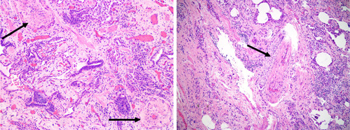
Figure 2.

Pulmonary vascular disease histology in interstitial lung disease. On the left panel, a histologic section of explanted lung tissue from a patient with idiopathic pulmonary fibrosis (IPF) who underwent lung transplant, showing marked concentric arterial medial hypertrophy (arrows) (Haematoxylin & Eosin, x100). On the right panel, explanted lung tissue of a patient with IPF without pulmonary hypertension (mean pulmonary artery pressure of 20 mmHg and pulmonary vascular resistance 1.5 wood units, pulmonary arterial wedge pressure 10 mmHg; data obtained 4 months before transplant) but showing signs of venous occlusion. With permission and many thanks to Dr. H. Mani (Inova Fairfax Hospital).
