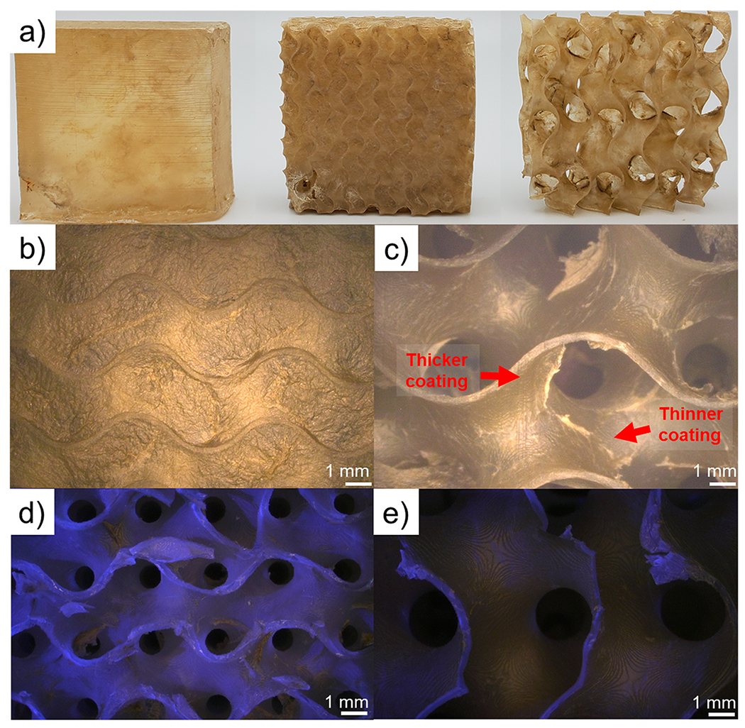
Figure 3.

Photographs of coated parts (a) from left to right: solid, small gyroid, and large gyroid. Optical micrographs of the exterior of a coated small gyroid, which has had its pores blocked by the thick growth of the nanobrick wall coating (b), exterior of a coated large gyroid (c). Optical micrographs under a combination of UV and white light illumination of: a cross-section of a coated small gyroid (d) and a cross-section of a coated large gyroid (e). Regions without fluorescence under UV light are attributed to the nanobrick wall coating. The large amount of fluorescence in part (d) highlights that very little coating penetrates to the interior of the small gyroid, while part (e) shows complete coverage of all surfaces.
