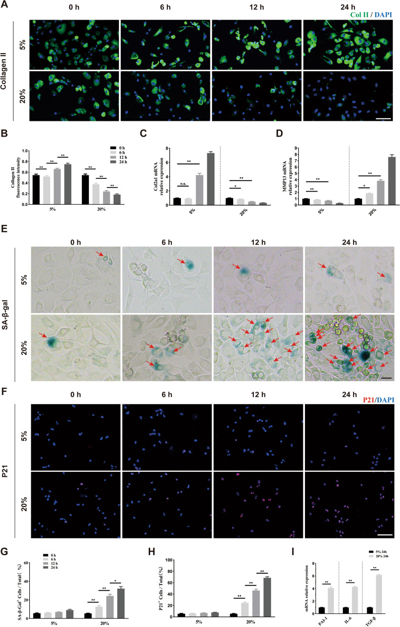
Fig. 2.
Excessive mechanical loading induces chondrocyte senescence in vitro. A, B Representative images and quantification of Collagen II staining (green) in primary chondrocytes treated with different elongation strain loading (5% or 20%) for 0, 6, 12, and 24 h. n = 6 per time point. Scale bar: 50 μm. C, D Quantitative PCR analysis of Col2a1 and Mmp13 in primary chondrocytes treated with different elongation strain loading (5% or 20%) for 0, 6, 12, and 24 h. n = 3 per time point. E, G Representative images and quantification of SA-β-Gal staining in primary chondrocytes treated with different elongation strain loading (5% or 20%) for 0, 6, 12, and 24 h. Red arrows represent SA-β-Gal.+ cells. n = 6 per time point. Scale bar: 20 μm. F, H Representative immunofluorescence images and quantification of P21 (red) in chondrocytes described in E. n = 6 per time point. Scale bar: 50 μm. I Quantitative PCR analysis of SASP-related genes (PAI-1, IL-6, TGF-β) in primary chondrocytes treated with different elongation strain loading (5% or 20%) for 24 h. n = 3 per group. All data are shown as the mean ± standard deviation (SD). *P < 0.05, **P < 0.01

