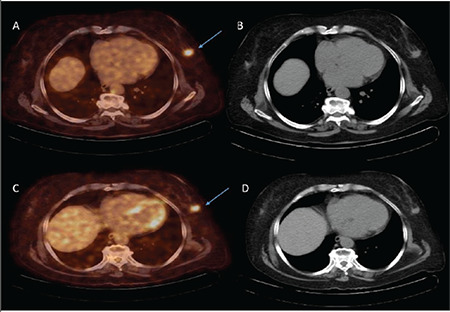
Figure 4.

Seventy-eight years old woman. Left breast localized invasive ductal carcinoma (primary tumor axial diameter 18.7 mm, primary tumor SUVmax:6.55) is seen in pretreatment CT and fusion PET/CT transaxial images (blue arrow) (A, B). There is a slight decrease in F-18 FDG uptake (SUVmax:4.51; ΔSUVmax:-44.25%) in posttreatment CT and fusion PET/CT transaxial images (blue arrow) after four cycles of cyclophosphamide/adriamycin chemotherapy (C, D). Histopathological features of the primary tumor: histological grade 2, nuclear grade 2, mitosis rate 2, ER 90% positive, PR 90% positive, HER2 negative, Ki-67 30%, p53 positive, and subtype luminal B/HER2 negative. Miller and Payne grading system pathological score 3
FDG: fluorodeoxyglucose; PET: positron emission tomography; CT: computerized tomography; SUV: standardized uptake value; HER2: human epidermal growth factor receptor 2; ER: estrogen receptor; PR: progesterone receptor
