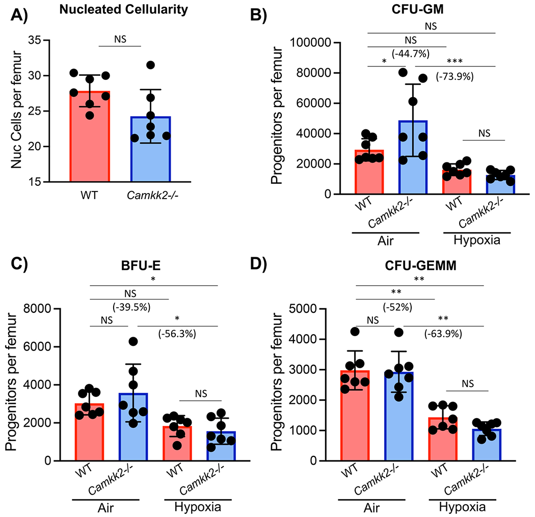
Fig. 3.

Nucleated cellularity and in vitro colony formation by wildtype (WT) and CaMkk2−/− bone marrow hematopoietic progenitor cells (HPC) isolated under hypoxia (3% O2) and then plated in ambient air (21% O2) versus hypoxia (3% O2). In a hypoxic glove box bone marrow from the femur of WT or KO mice was harvested in sterile PBS, counted, and split in half so that one half remained under hypoxia and the other half was acclimated to ambient air for 2 hours. Nucleated cells were counted and methylcellulose cultures established to enumerate CFU-GM, BFU-E, and CFU-GEMM. Cells were plated at 5 × 10⁴ nucleated cells per ml containing fetal bovine serum (30% v/v), PWMSCM (5% vol/vol), mSCF (50 ng/ml), erythropoietin (1 U/ml), and Hemin (0.1 mM). Cultures were incubated for 6 days at 5%CO2/5% oxygen in a humidified environment. All culture reagents used to plate cells remaining in hypoxia were acclimated in the hypoxia chamber for 16-18 h. Results are shown for 2 separate experiments with a total n = 7. (A) Nucleated cellularity, (B) CFU-GM, (C), BFU-E, and (D) CFU-GEMM are shown. Statistical analysis was performed using t-tests (A) or two-way ANOVA with Tukey post hoc tests (B-D). *p < 0.05; **p < 0.01; ***p < 0.001
