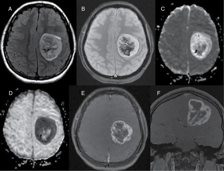
Figure 1.
Preoperative images. (A) Axial FLAIR image shows 5 cm mass centered in the left centrum semiovale and corona radiata with surrounding minimal vasogenic edema. (B) On T2 GRE, there is central susceptibility compatible with internal hemorrhage. (C) On diffusion weighted image, the peripheral components show mostly isointense signal with increased signal in the lateral margin. Internal heterogeneity, likely due to internal blood products. (D) ADC map shows corresponding bright signal in the peripheral regions with dark signal in the lateral margin, suggestive of facilitated diffusion and low cellularity mostly with relative diffusion restriction in the lateral margin. Heterogeneous signal at the center was due to blood products. Fat-saturated contrast enhanced axial (E) and coronal (F) images show avid thick enhancement in the peripheral solid component with irregular lobulated contours and bell-pepper like appearance. Overall, MRI features are suggestive of an aggressive high-grade glial tumor except the diffusion weighted images, demonstrating facilitated diffusion in the most of the solid peripheral components with small focal diffusion restriction in the lateral margin.

