Figure 8.
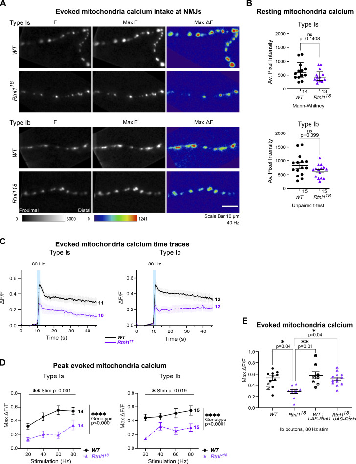
Rtnl1 loss decreases evoked mitochondrial Ca2+ uptake in Ib and Is presynaptic termini. Evoked mitochondrial Ca2+ responses were measured at Type Ib and Type Is termini at muscle 1 in segment A4-A6. (A) Mitochondrial CEPIA3mt fluorescence presented as in Fig. 6 A. (B) Loss of Rtnl1 does not alter resting mitochondrial Ca2+ levels in Type Is or Type Ib terminals. Some outlier datapoints are excluded from the graph but included in statistics. Graphs and analyses are as in Fig. 6 B, except that Type Ib are shown as mean ± SEM and compared using Student’s t test. (C) Time courses of evoked mitochondrial CEPIA3mt responses to 80 Hz stimulation (mean ± SEM) show reduced Ca2+ uptake by mitochondria on loss of Rtnl1. (D) Loss of Rtnl1 significantly decreases mitochondrial Ca2+ uptake across a range of stimulation frequencies. Graphs and analyses are as for Fig. 5 D. (A–D) Genotypes: Is-GAL4 or Ib-GAL4, UAS-CEPIA3mt/UAS-tdTom::Sec61β in either a WT or Rtnl118 mutant background. (E) Expression of UAS-Rtnl1::HA under Ib-GAL4 control rescues evoked Ca2+ uptake by mitochondria in mutant larvae. Data shows maximum evoked mitochondrial CEPIA3mt responses (ΔF/F) after 80 Hz stimulation. Graph shows individual larval datapoints, with one NMJ datapoint per larvae, averaged across the responding area; statistical comparisons were performed using one-way ANOVA followed by Dunnett's post-hoc multiple comparisons. Genotypes: Ib-GAL4, UAS-CEPIA3mt/UAS-Rtnl1::HA or Ib-GAL4, UAS-CEPIA3mt/attP2+ in either a WT or Rtnl118 mutant background; the attP2 landing site acts as a WT control for insertion of UAS-Rtnl1::HA at attP2.

