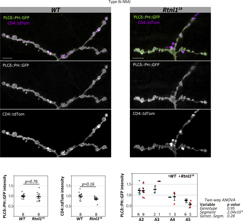
Figure S6.
Rtnl1 loss does not affect presynaptic PI(4,5)P2 levels. Representative examples of confocal projections and quantifications of WT and Rtnl118 larvae showing the distribution of the PI(4,5)P2 marker PLCδ::PH::GFP in Type Ib muscle 1 NMJ. Scale bars, 10 μm. Plots show individual larval datapoints and mean ± SEM; y-axis indicates arbitrary units (au) after normalization to control (WT); sample size (larvae) is indicated within the plot for each genotype. For each larva, several NMJs between A2-A6 segments were analyzed, and the mean larval value is shown as a datapoint. Student’s t tests were performed for pairwise comparisons. Genotypes are Ib-GAL4, UAS-CD4::tdTom/UAS-PLCδ::PH::GFP, in either a Rtnl1+ (WT) or Rtnl118 background.

