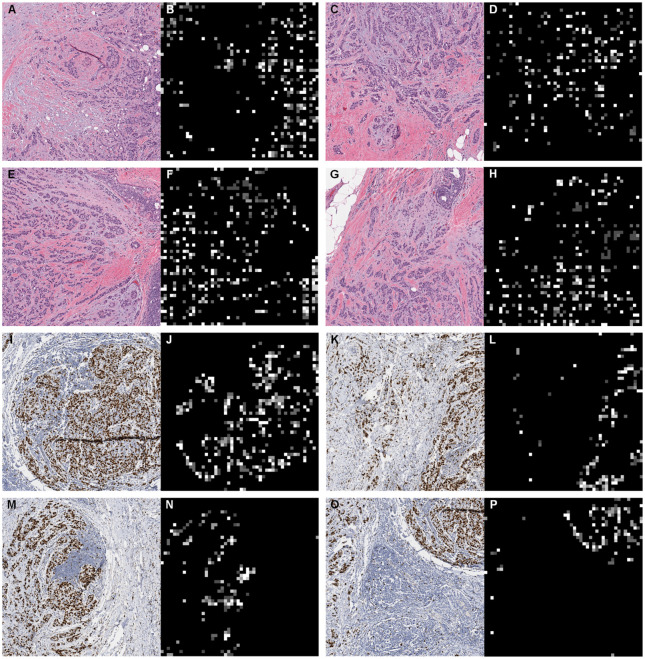
Fig 11. Magnified local regions and corresponding heatmaps.
We can clearly find that the attention based MIL is highlighting specific tissue patterns. This is especially interpretable on Ki67 images, where proliferating cells (i.e. brown color regions on WSI images) are assigned with high attention weights (i.e. bright regions on heatmaps).

