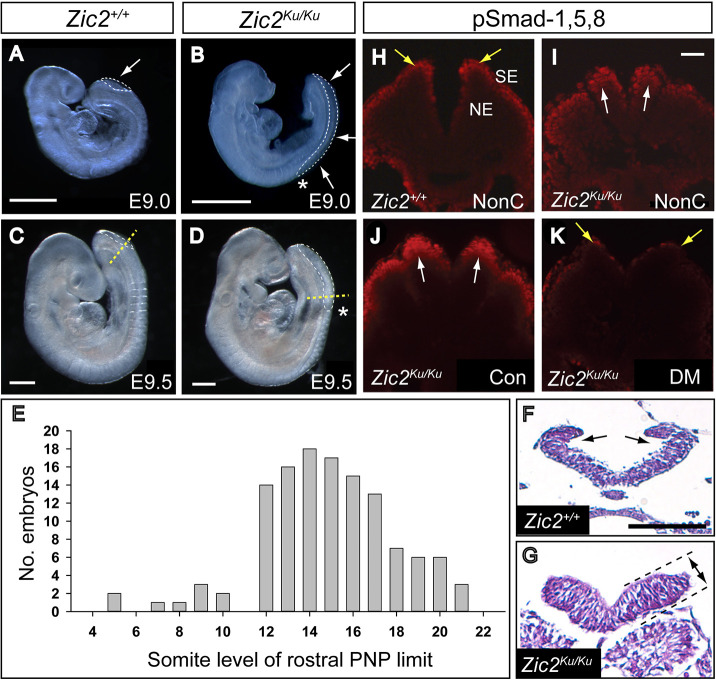
Fig. 1.
Spinal neurulation failure and enhanced pSmad-1,5,8 expression in Zic2Ku/Ku mutants. (A-D) Defective spinal closure, as shown by the enlarged posterior neuropore (PNP) (outlined by white dashed lines; arrows in A,B), in Zic2Ku/Ku embryos at E9.0 (B) and E9.5 (D) compared with normally developing wild-type (Zic2+/+) littermates (A,C). (E) Somite level of the rostral limit of the PNP (asterisks in B,D) among 124 Zic2Ku/Ku embryos at E9.5-10.5. Closure does not usually progress beyond the level of somites 12-17, at which level dorsolateral hinge points (DLHPs) first appear in normal development. (F,G) Transverse sections (Haematoxylin and Eosin-stained) through the rostral PNP of E9.5 Zic2+/+ (F) and Zic2Ku/Ku (G) embryos, at the level of the yellow dashed lines in C,D, respectively. Both show equivalent neural fold elevation, but paired DLHPs are present in the Zic2+/+ embryo (arrows in F) and absent from the Zic2Ku/Ku embryo (G). The mutant neuroepithelium shows marked apicobasal thickening (double-headed arrow between dashed lines in G). (H-K) Immunohistochemistry for pSmad-1,5,8 on transverse sections through the elevated neural folds of Zic2+/+ (H) and Zic2Ku/Ku (I-K) embryos. In non-cultured (NonC) 15-somite-stage embryos (H,I), immunoreactivity is enhanced in the dorsal Zic2Ku/Ku neuroepithelium (arrows in I), compared with that in Zic2+/+ embryos, in which expression is mainly in surface ectoderm (arrows in H). Zic2Ku/Ku embryos exposed for 16 h in culture to vehicle (Con) from E8.5 show pSmad-1,5,8 immunoreactivity comparable to that seen in non-cultured Zic2Ku/Ku embryos (compare J with I). Dorsomorphin exposure in culture produces markedly reduced pSmad-1,5,8 expression (K), similar to that in non-cultured Zic2+/+ embryos. NE, neuroepithelium; SE, surface ectoderm. Scale bars: 0.5 mm (A-D); 50 µm (F); 50 µm (I).

