Fig. 2.
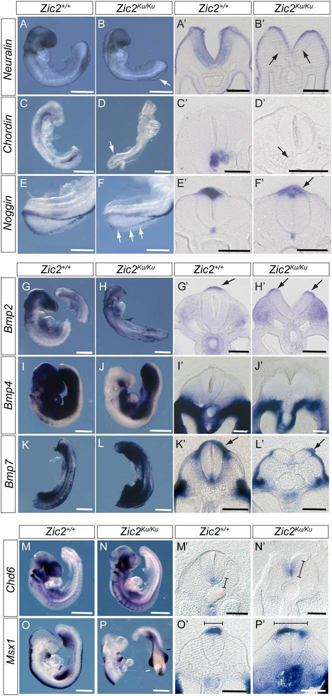
Overactivation of BMP signalling in pre-spina bifida Zic2Ku/Ku embryos. (A-P′) Comparison of Zic2+/+ (A,A′,C,C′,E,E′,G,G′,I,I′,K,K′,M,M′,O,O′) and Zic2Ku/Ku (B,B′,D,D′,F,F′,H,H′,J,J′,L,L′,N,N′,P,P′) embryos at E9.5 for expression of BMP antagonists (A-F′), BMP ligands (G-L′) and downstream markers of BMP signalling (M-P′). Whole-mount in situ hybridisation was followed by vibratome sectioning through the closing or recently closed spinal neural tube. (A-F′) BMP antagonists neuralin, chordin and noggin are expressed on the tips of the neural folds throughout the PNP, and all show reduced expression in Zic2Ku/Ku embryos compared with wild-type littermates (arrows in B,B′,D,D′,F,F′). (G-L′) In contrast, Bmp2 and 7 show no marked differences in dorsal expression intensity between genotypes (arrows in G′,H′,K′,L′), while Bmp4 is not expressed dorsally (I′,J′). Apparent increase in cranial expression of Bmp4 in the wild-type embryo (I) is due to probe trapping. (M-P′) Domains of Chd6 and Msx1 expression in the neural tube, which are regulated by BMP signalling, are extended in Zic2Ku/Ku embryos compared with wild-type embryos (lines in M′,N′,O′,P′). Strong signal for Msx1 in the gut of the mutant embryo (P′) is due to probe trapping. Three embryos were hybridised for each probe and genotype combination except for Bmp2 where n=2, with representative images shown. Scale bars: 0.5 mm (A-D,G-P); 1 mm (E,F); 50 µm (A′-L′); 100 µm (M′-P′).

