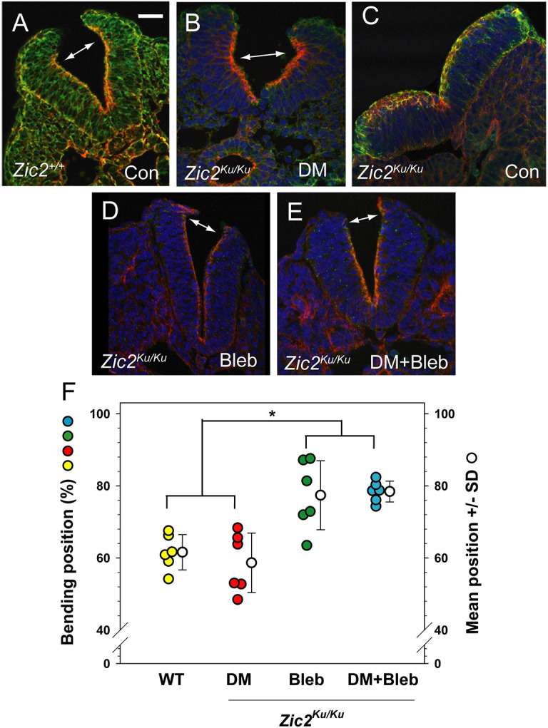
Fig. 4.

Neural plate bending at distinct ventrodorsal positions after partial rescue of closure by DM or Bleb. (A-E) Zic2+/+(A) and Zic2Ku/Ku (B-E) embryos at the 15- to 19-somite stage, either uncultured (A,C; Con) or cultured in the presence of DM, Bleb or both (B,D,E). Transverse sections at rostral PNP level are stained with Phalloidin (red), anti-MHC (green) and DAPI (blue). Note the typical DLHPs in the wild-type neural plate (double arrow in A), and closely similar bending points in the DM-treated mutant neural plate (double arrow in B). An untreated mutant (C) shows a complete lack of DLHPs and an apicobasally thickened neural plate. Zic2Ku/Ku embryos treated with Bleb or Bleb+DM show more distally located bending, just below the dorsal neural plate tips (double arrows in D,E). (F) Measurement of bending position, as % of ventral–dorsal (V-D) distance along the apical neural plate border. Wild-type and DM-treated mutant embryos exhibit bends clustering ∼60% of the V-D distance whereas Bleb (+/− DM) induces bends clustering ∼80% of the V-D distance (*P=0.002; one-way ANOVA with post-hoc Holm-Sidak tests). Three embryos (two neural folds per embryo) measured in each group. Scale bar: 30 µm.
