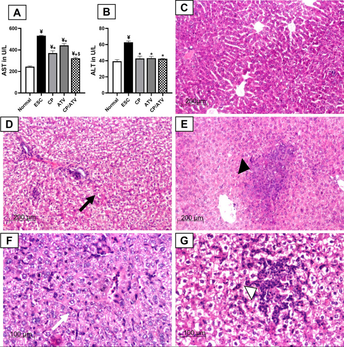
Fig. 1.
Hepatic transaminases and representative photomicrographs of H&E-stained liver sections: mean level of serum AST and ALT in A, B, respectively. ¥p < 0.05 versus normal control; *p < 0.05 versus ESC control; $p < 0.05 versus ATV-treated group. AST: Aspartate aminotransferase; ALT: Alanine aminotransferase; Normal: normal control; ESC: Ehrlich solid carcinoma control group; CP: cyclophosphamide-treated ESC group; ATV: Autoclaved Toxoplasma vaccine-treated ESC group; CP/ATV: Combined cyclophosphamide-treated and ATV-treated ESC group. Hepatic H&E sections showed: C normal preserved architecture in normal control. D Diffuse fatty changes (black arrow) with mild inflammation in ESC control. E Focal fatty changes and scattered granuloma composed of inflammatory cells (black arrowhead) in CP-treated group. F Moderate inflammatory cells aggregate mainly lymphocytes especially in the sinusoidal spaces with Kupffer cell hypertrophy (white arrow) in ATV-treated group. G Mild to moderate degree of portal inflammation with foci of lymphocytic aggregates and focal necrosis, marked hypertrophy and hyperplasia of Kupffer cells and sinusoidal lymphocytic infiltration (white arrowhead) in CP/ATV-treated group

