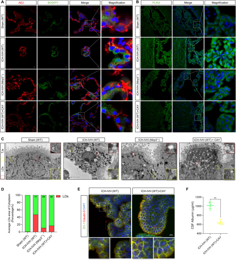
Fig. 5. Inhibiting LD formation improved B-CSFB permeability and neurological deficits after ICH-IVH.
A BODIPY staining of LDs in the choroid plexus (marker with AE2). Scale bar = 20 μm. B Representative images of PLIN3 staining indicating LDs in the choroid plexus. Scale bar = 50 μm. C TEM images showed tight junctions and LDs in the choroid plexus. Scale bar = 1 μm. D LD area percentage of total cytoplasm according to TEM images. **P < 0.01, sham (WT) versus ICH-IVH (WT); ##P < 0.01, ICH-IVH (WT) versus others (n = 3, one-way ANOVA). E Immunofluorescence staining of ZO-1 and claudin-5 in the choroid plexus after inhibiting LDs. Scale bar = 20 μm. F CSF albumin content after ICH-IVH with or without CAY10650 treatment (n = 6, unpaired t test). The results are expressed as the mean ± SD, **P < 0.01.

