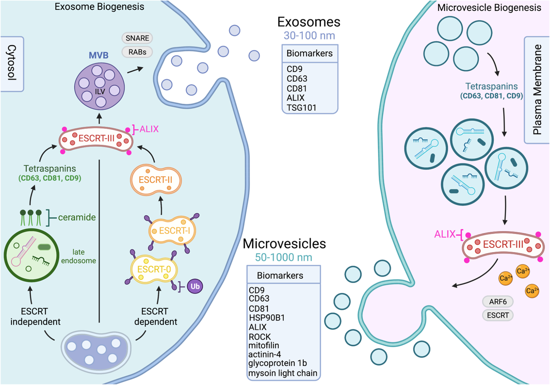
Figure 2. Extracellular vesicles biogenesis occurs by invagination or blebbing on the cell membrane originating different vesicle subtypes.

Extracellular vesicle biogenesis differs depending on the sub-population of vesicles in question. Exosomes range from 30–100nm in size, formed by plasma membrane invagination utilizing ESCRT-independent, shown to include ceramides and tetraspanin driven sorting, and ESCRT-dependent pathways, which gathers ubiquitylated cargo using ESCRT protein complexes. The release of exosomes derived from both pathways are influenced by SNARE and Ras-related proteins. Microvesicles are generally larger, ranging from 50–1000nm in size, and form through blebbing and budding on the plasma membrane. Microvesicles share components with exosome formation, including tetraspanins, however cargo in this case is determined by a component’s lipid raft affinity and anchorage to plasma membrane. MVBs fuse with the plasma membrane and release the ILVs into the extracellular space. The budding and consequential release of microvesicles is influenced by Ca2+ levels, ESCRT pathway associated proteins, and ADP-ribosylation factor 6. Exosomes and microvesicles have distinct surface markers to help differentiate the two populations from each other.
