Figure 1.
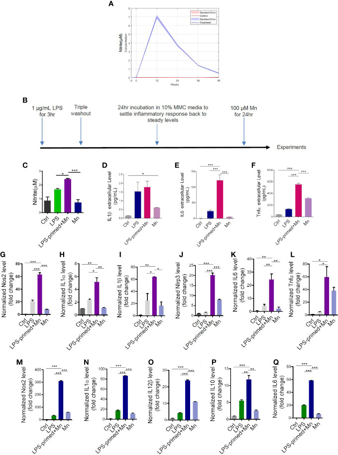
Heightened inflammatory responses in trained immunity. (A) LPS-induced time response experiment in mouse microglial cells (MMCs) via Griess assay. Data are means ± SEM of two individual experiments performed in four replicates. Light blue area denotes SEM. (C) Nitric oxide release from LPS- and Mn-treated, primed MMC via Griess assay (n=8-9), following the treatment paradigm in (B). LPS+Mn denotes the treatment with LPS for 3 h, a 24-hour recovery, and then Mn for 24 h. (D–F) Luminex bead-based cytokine assays (n=3) of pro-inflammatory cytokine release following the treatment paradigm in (B). (G–L) RT-qPCR analysis of Nos2, IL-1α, IL-1β, NLRP3, IL-6, and Tnfα in Mn-exposed, LPS-primed PMG cells following the treatment paradigm in (B). Two individual experiments (n=2) were performed. (M–Q) RT-qPCR analysis of Nos2, IL-1α, IL-12β, IL-10, and IL-6 in trained MMC, using the treatment paradigm in (B). Two individual experiments (n=2) were performed. Data show mean ± SEM of one-way ANOVA followed by Tukey’s post hoc test. Ctrl, control; *p ≤ 0.05; **p<0.01; ***p<0.001.

