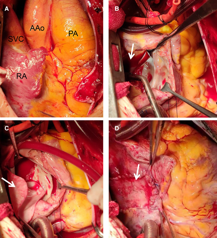
Figure 3.
Intraoperative view of repair procedure: during the operation, the right atrium, right ventricle, pulmonary artery and superior vena cava were found significantly enlarged (A); A double-chamber incision was made and the posterior wall of the left atrium was opened (B, arrow); the common pulmonary veins were anastomosed with the posterior wall of the left atrium. A proper size of the bovine pericardium patch (C, arrow) was used to close the ASD and enlarged the left atrium. A proper size of autologous pericardium was used to close the right atrial incision (D, arrow). AAo, ascending aorta; PA, pumonary artery; RA, right atrium; SVC, superior vena cava.

