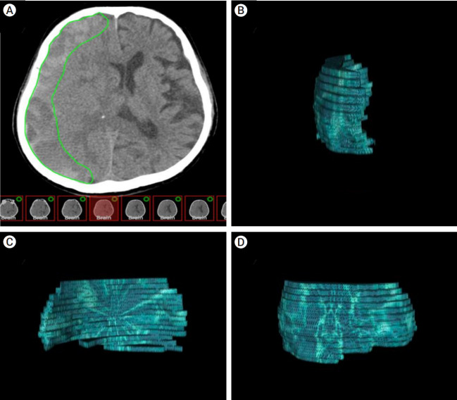
Fig. 1.
The hematoma model was reconstructed using OsiriX software (an open-source DICOM viewer, OsiriX Lite, v.12.5.2 32bit, Pixmeo, Geneva, Switzerland). (A) Axial CT-scan showing a right-sided subdural hematoma (SDH). Volumetric measurements were performed by hand-tracing the hematoma form in each slice, as shown in green. (B) 3D frontal aspect reconstruction of computer-assisted volumetric measurement of single-sided SDH. (C) 3D medial aspect reconstruction of computer-assisted volumetric measurement of single-sided SDH. (D) 3D lateral aspect reconstruction of computer-assisted volumetric measurement of single-sided SDH. CT, computed tomography

