Figure 3.

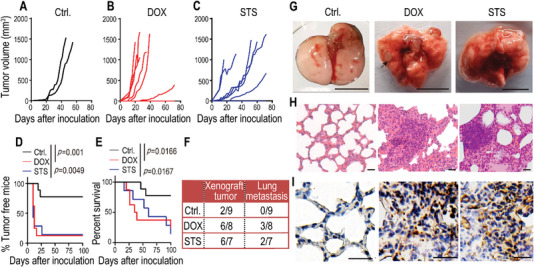
Exacerbated metastatic capability of NDCs in a xenograft model. A–C) 8 × 10⁵ luciferase‐labeled HeLa cells (HeLa‐Luc, Ctrl.) and DOX‐ or STS‐induced HeLa‐Luc NDCs were subcutaneously inoculated into female BALB/c‐nude mice. A) Xenograft tumors occurred in mice with HeLa‐Luc (n = 9), B) NDCs derived from DOX‐treated HeLa‐Luc (n = 8), and C) STS‐treated HeLa‐Luc (n = 7). D) Tumor‐free survival and E) overall survival of mice from the above experiments (A–C). p values were determined by a Log‐rank t test. F) The lung metastasis rates of HeLa‐Luc xenograft mice from the above experiments (A–C) on day 72 after inoculation. G) Gross photography showing apparent pulmonary metastases in xenograft mice with DOX‐ and STS‐induced HeLa‐Luc NDCs. The scale bars represent 1 cm. The black arrow indicates the site of the tumor nodule. H) H&E staining and I) IHC staining (anti‐luciferase antibody) of lung sections that show obvious metastatic masses in DOX‐ and STS‐induced HeLa‐Luc NDCs. The scale bars represent 50 µm for (H), and 20 µm for (I).
