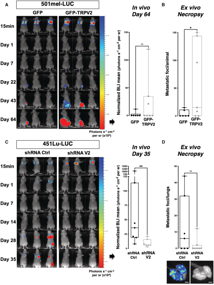
Figure 6. TRPV2 expression level dictates the in vivo metastatic potential of human melanoma tumor cells xenografted in mice.

-
A–DRepresentative bioluminescence imaging (BLI) data of mice injected intravenously with the nonmetastatic melanoma cell line 501mel‐Luc transfected with either GFP‐TRPV2 or GFP control (A, B), or with the invasive 451Lu‐Luc melanoma cell line expressing either control shRNA or TRPV2 targeting shRNA (C, D). Tumor growth and metastasis formation were monitored for 64 days (501mel‐Luc) or 35 days (451Lu‐Luc) after injection. Graphs presented in A and C show in vivo normalized photon flux quantification at the end time point (ns P = 0.0903 (A) and **P = 0.0064 (C), the Mann–Whitney test). Graphs presented in (B) and (D) show the number of metastatic foci per animal (B) or per lungs (D) counted at necropsy (*P = 0.0397 (B) and ns P = 0.0691 (D), unpaired t‐tests). For (D) Representative ex vivo BLI images of lung metastasis are shown (Scale bar = 5 mm).
Data information: Boxes extend from the 25th to 75th percentiles, whiskers from min to max, the horizontal line in each box is plotted at the median and each dot correspond to a single mouse (n = 6–7).
Source data are available online for this figure.
