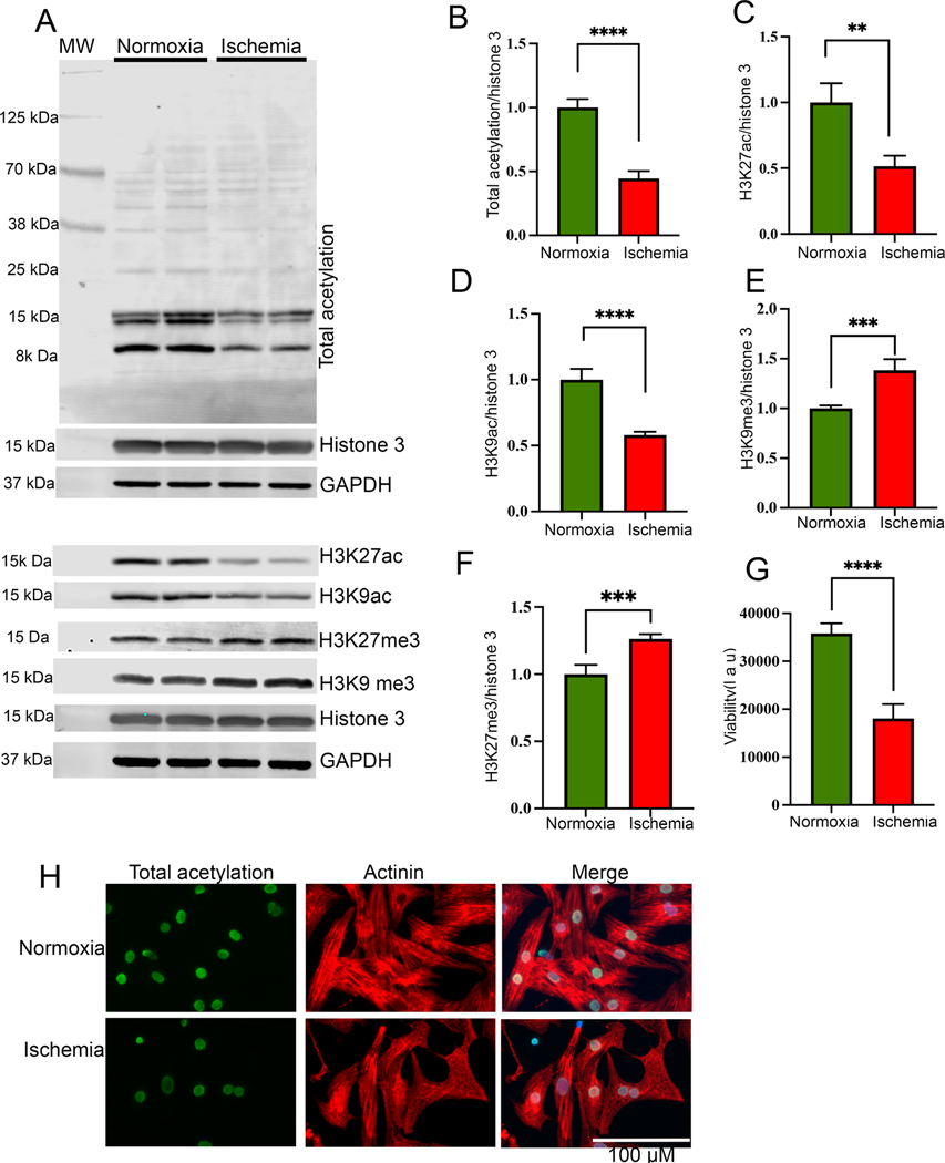
Figure 1:
Histone acetylation significantly reduced during ischemia. (A-F) Western blots show the cellular acetylation and methylation during normoxia and ischemic condition. NRVCs were incubated in normoxia and ischemic condition for 24 h and western blots were performed using total protein lysate and blots were probed with H3K27ac, H3K9ac, total acetylation, H3K27me3 H3K9me3, GAPDH and histone antibodies. Graphs show the quantification data of the western blot (**, p value<0.05; ***, p value<0.05; ****, p value<0.0001). (G) The graph shows viability of NRVCs after exposing them to normoxic and ischemic conditions (****, p value<0.0001). (H) Representative images show acetylation of NRVCs exposed to normoxia and ischemic condition for 24 h. Cells were fixed with 4% PFA and immunocytochemistry were performed with total acetylation (green) and actinin (red) antibodies and nucleus were stained with DAPI (blue). Images of fixed cells were captured by Keyence microscope and analyzed by BZ-X800 analyzer (n=50 cells in each condition).

