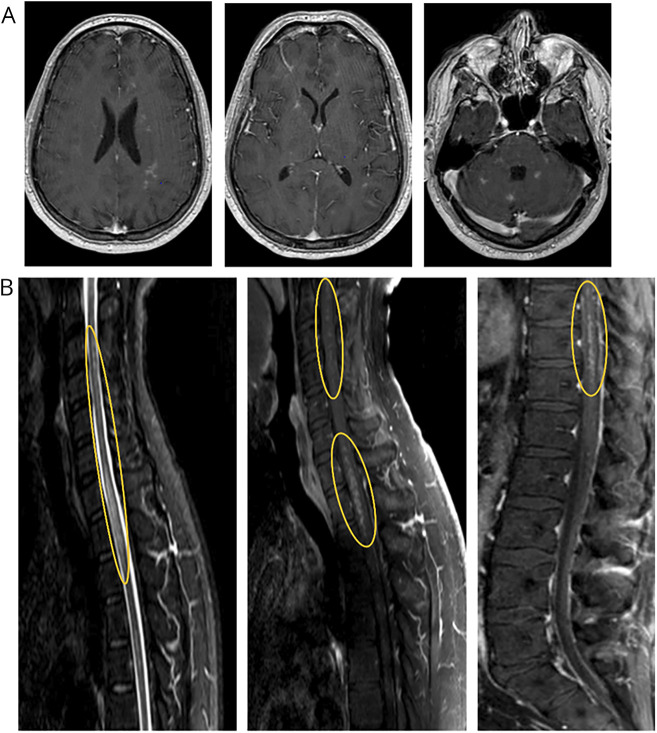
Figure 1. Pretreatment MRI Demonstrating Multifocal Enhancing Lesions.
(A) Contrast-enhanced brain MRI demonstrating punctate enhancing lesions scattered throughout the brain parenchyma including the subcortical white matter, basal ganglia, cerebellum, and brainstem. (B) Highlighted in the yellow ovals are the prominent areas of punctate, perivascular appearing enhancement throughout the spinal cord.

