
FIGURE 3.
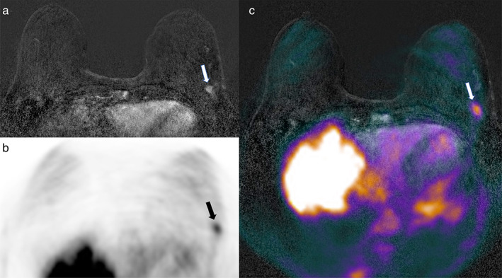
Axial (a) subtracted dynamic contrast‐enhanced (DCE)‐MRI, (b) PET, and (c) fused DCE‐MRI and PET imaging. A 66‐year‐old patient with invasive ductal breast cancer (9 mm, G3, ER/PgR−, HER2+) of the left breast (arrows in a–c).
Official websites use .gov
A
.gov website belongs to an official
government organization in the United States.
Secure .gov websites use HTTPS
A lock (
) or https:// means you've safely
connected to the .gov website. Share sensitive
information only on official, secure websites.
Axial (a) subtracted dynamic contrast‐enhanced (DCE)‐MRI, (b) PET, and (c) fused DCE‐MRI and PET imaging. A 66‐year‐old patient with invasive ductal breast cancer (9 mm, G3, ER/PgR−, HER2+) of the left breast (arrows in a–c).