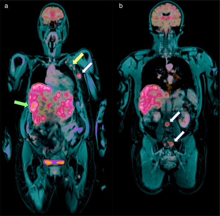
FIGURE 5.

(a and b) Fused PET and post‐contrast fat‐saturated T1‐weighted imaging on the coronal plane (whole‐body examination) shows liver and axillary involvement (green and yellow arrows in a, respectively) as well as rib and lumbo‐sacral bone metastases (white arrows in a and b, respectively) in in a 66‐year‐old patient with invasive ductal breast cancer (G3, ER/PgR−, HER2+) in the left breast (same patient as the patient in Figs. 3 and 4).
