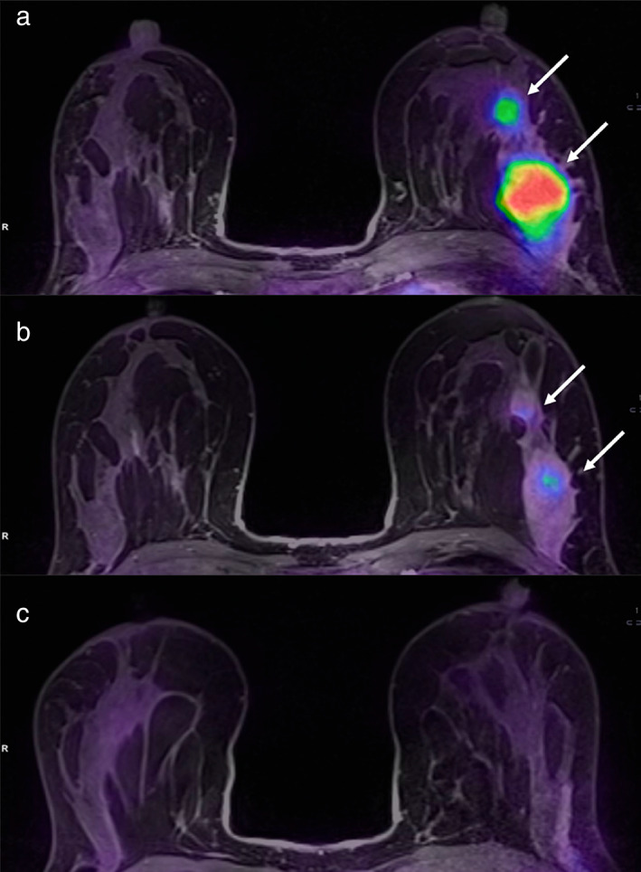
FIGURE 15.

A 36‐year‐old patient with left breast cancer undergoing NAC. Fused PET/MRI images acquired before (a), during (b), and after (c) NAC are shown. While a slight reduction of the tumor and its satellite nodule (white arrows in b) is appreciable, 18FFDG uptake is significantly reduced after the second cycle of chemotherapy (b) as compared to the pre‐treatment evaluation (a). The tumor was not detectable at the post‐treatment evaluation (c). Pathology after surgery demonstrated a complete response. Reprinted under a Creative Commons (CC BY 4.0) license from: Romeo V, Accardo G, Perillo T, Basso L, Garbino N, Nicolai E, Maurea S, Salvatore M. Assessment and Prediction of Response to Neoadjuvant Chemotherapy in Breast Cancer: A Comparison of Imaging Modalities and Future Perspectives. Cancers (Basel). 2021;13(14):3521.
