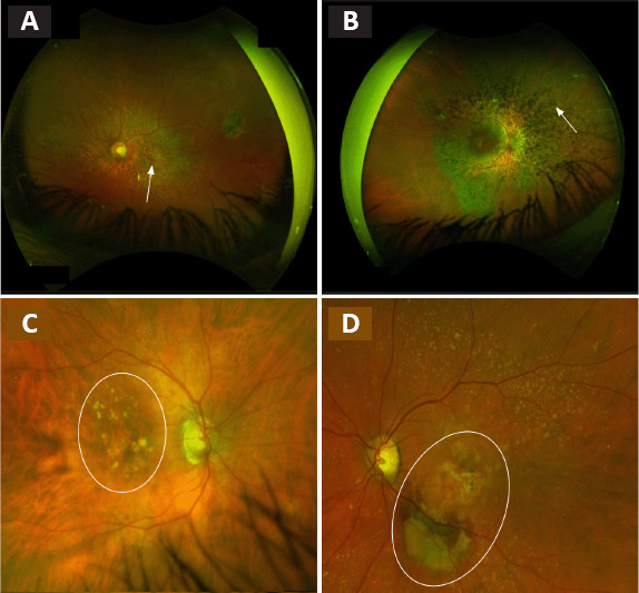
Figure 1.

Fundus examination of patients affected by retinal diseases.
(A–D) STGD1 disease with the atrophy of the macular area (white arrow) (A), RP with the out bone spicule pigmentation (white arrow) (B), dry AMD with drusen and atrophy within the macular area (surrounded in white) (C) or wet AMD with fibrosis and retinal hemorrhage (surrounded in white) (D). AMD: Age-related macular degeneration; RP: retinit pigmentosa; STGD1: Stargardt macular degeneration. Unpublished data.
