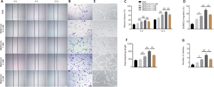
Figure 4.
Metformin promotes SCMEC-mediated angiogenesis in vitro.
(A) Representative images of wound-healing experiment by SCMECs treated with different doses of metformin or PBS. Metformin markedly increased wound healing by SCMECs, (B) Representative images of the results from a Transwell experiment in which SCMECs were treated with different doses of metformin or PBS. Metformin dramatically increased SCMEC migration. (C) Quantification of the wound closure results shown in A. (D) Quantification of the SCMEC migration results shown in B. (E) Representative images of tube formation by SCMECs treated with different doses of metformin or PBS. A marked increase in tube formation by SCMECs was observed after administration of different doses of metformin. Scale bars: 300 μm in A, 200 μm in B, 100 μm in E. (F, G) Quantification of the total branching length and tube mesh extension data shown in E. The data are presented as the mean ± SEM (n = 6 per group). #P < 0.05, ##P < 0.01 (one-way analysis of variance followed by Tukey’s multiple comparisons test). PBS: Phosphate buffer saline; SCMECs: spinal cord microvascular endothelial cells.

