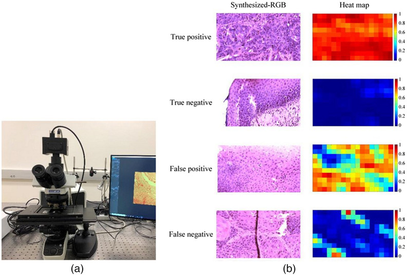
Fig. 12.
(a) A compact hyperspectral Snapscan camera on top of a microscopic system (reproduced from Ref. 47). (b) Using the system described, a series of hyperspectral digital pathology images were acquired. The slides were from patients diagnosed with squamous head and neck carcinoma. A machine learning system was used to produce a probability heat map of cancer occurrence (reproduced from Ref. 240).

