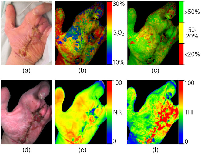
Fig. 13.
Estimation of physiological values from a wound photograph. (a) RGB image, (b) and (c) relative and segmented tissue oxygenation mapping ( in the figure), (d) reconstructed RGB image from hyperspectral data, (e) near-infrared-based perfusion data (NIR in the figure), and (f) relative tissue hemoglobin index (THI in the figure) (reproduced from Ref. 257).

