Fig. 2.
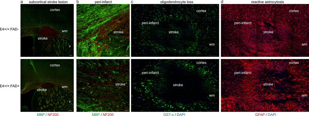
Subcortical stroke in human ApoE4-TR:5XFAD transgenic mice. Immunolabeling for myelin basic protein (green) and neurofilament-200 (NF200, red) at 7 days after stroke demonstrates a similar size-targeted ischemic lesion in the subcortical white matter in both E4-TR:FAD- (top) and E4-TR:FAD+ (bottom) animals (a). Dense loss of myelin and axonal swellings are present within the stroke core and peri-infarct white matter (b). No difference was observed in oligodendrocyte loss (c) or reactive astrocytosis (d) comparing stroke lesions in E4-TR:FAD- (top) and E4-TR:FAD+ (bottom) animals. Scale bars = 200 μm (a), 50 μm (b), 100 μm (c, d); wm, white matter; v, ventricle

