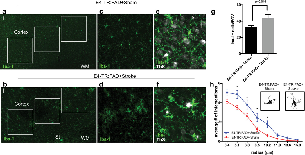
Fig. 5.
Microglial analysis in human ApoE4-TR:5XFAD transgenic mice with subcortical stroke. Immunostaining for Iba-1 in the cortex of 5-month-old E4-TR:FAD+ mice 3 months after the sham (a, c) or stroke (b, d) procedure. Regions of interest (white boxes) in the ipsilateral cortex were used to quantify the number of Iba1+ cells (a, b). Co-labeling with Iba1 (green) and thioflavin S (ThS, white) shows the localization of plaques in relation to Iba1+ cells (e, f). Quantification of the number of Iba1-positive cells per field of view (g). Microglial morphology measured using Sholl analysis (n = 45 cells/animal) demonstrates an increase in activated microglial branching between E4-TR:FAD+ sham and E4-TR:FAD+ stroke (p < 0.0001 by two-way ANOVA, F = 32.09; *adjusted p < 0.05) (h). Insets show binary masked examples of microglial morphology from each condition. Scale bars = 50 μm in (a) and (b); 20 μm in (c)–(f). St, stroke; WM, white matter; Ve, ventricle

