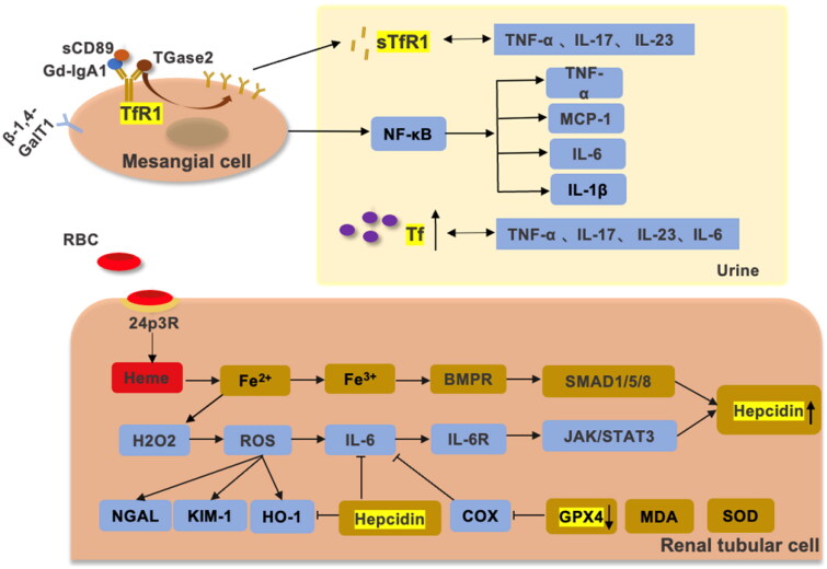
Figure 3.
The relationship between iron metabolism, IgAN, and chronic inflammation. β-1,4-GalT, sTfR1, and CD89 are expressed by antibodies on mesangial cells in IgAN. Gd-IgA1 activates inflammation by promoting TfR1 overexpression through positive feedback to promote the secretion of IL-6, TNF-α, IL-1β and MCP-1 by the NF-κB pathway [107–109]. Overexpressed TfR1 can release sTfR1, which positively correlates with TNF-α, IL-17 and IL-23. When the glomerular filtration barrier is disrupted, Tf is increased in the urine. Tf is positively correlated with TNF-α, IL-17, IL-23, IL-6 and CRP. Ferroptosis is present in IgAN, and hemoglobin from hematuria is reabsorbed by 24p3R in renal tubular cells. Due to the tubular toxicity of hemoglobin, Fe3+ produced by the Fenton reaction promotes oxidative stress, causing increased expression of NGAL, KIM-1, HO1 and IL-6. The SMAD/BMP pathway and JAK/STAT3 pathway promote the expression of hepcidin [11,110]. GPX4 and SOD expression was decreased and MDA expression was increased in IgAN. GPX4 can suppress IL-6 by inhibiting LOX. In addition, hepcidin achieves renal protection by inhibiting IL-6 and HO-1 in hemoglobin-mediated renal tubular injury.

