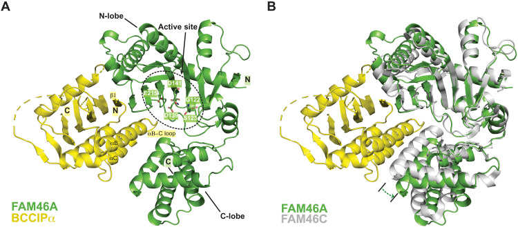
Fig. 3. Crystal structure of the FAM46A/BCCIPα complex.
(A) Overall structure of the FAM46A/BCCIPα-ΔL complex. Residues in the active site of FAM46A are highlighted. The structural elements in BCCIPα involved in binding to FAM46A are labeled. N and C denote the N and C termini of the proteins, respectively. (B) Structural comparison between FAM46A in the FAM46A/BCCIPα complex and FAM46C in the apo-state (PDB ID: 6W36). The structures are superimposed on the basis of the N-lobe of FAM46A and FAM46C. The dashed arrow indicates that the C-lobe of FAM46A in the complex rotates away from the N-lobe compared with that in apo-FAM46C.

