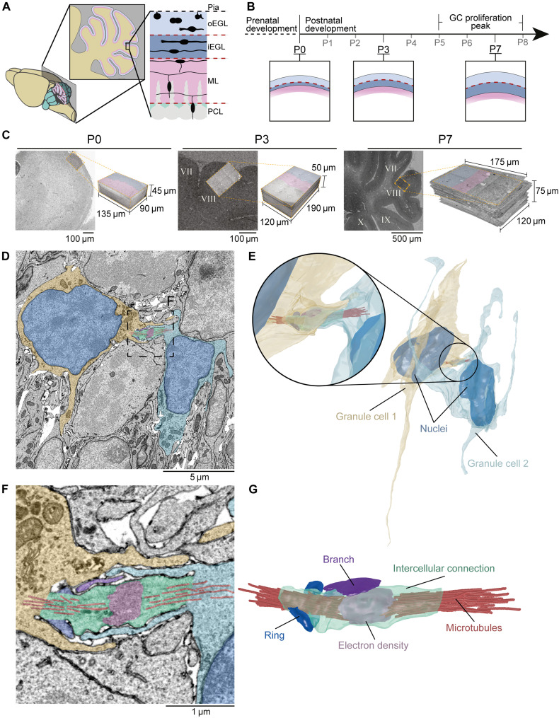
Fig. 1. Identification of cerebellar GC intercellular connectivity.
(A) Schematic representation of a mouse cerebellar cortex sagittal cross section. Sagittal view of the three outer laminae of the cerebellar cortex: the EGL (blue) divided into outer and inner EGL (oEGL and iEGL), the molecular layer (ML; pink), and Purkinje cell layer (PCL; green). (B) During pre- and postnatal development, GCs populate the oEGL via mitosis, resulting in a sequential expansion of the EGL (P0, P3, and P7 inserts) and a proliferation peak between postnatal days 5 and 8 (P5 and P8). Postmitotic GCs arising from these divisions then migrate tangentially in the iEGL. To become mature neurons, postmitotic GCs migrate radially through the ML and PCL and settle in the internal granular layer (IGL), where they establish synapses with Golgi cells and Mossy fibers (not shown). (C) P0, P3, and P7 electron micrographs and 3D volumes prepared using serial-sectioning scanning electron microscopy (ssSEM); lobule VIII for P3 and P7 volumes. (D) 2D electron micrograph showing GCs (yellow and blue) in the EGL of the P7 volume bridged by an intercellular connection (IC; green). (E) 3D reconstructions of D. (F and G) Zoomed-in micrograph (F) and 3D reconstruction (G) of (D) and (E), respectively, showing microtubules (red) that emanate into both GCs, an electron-dense region at the center of the IC (pink), an IC branch (magenta), and an incomplete ring (blue) extruding from the IC.

