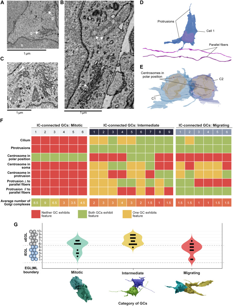
Fig. 4. IC-connected cells are mitotic, migrating, or transitioning between the two states (intermediate).
(A to C) Identification of cellular organelles within GCs in electron micrographs: n, nucleus; c, centrosome; g, Golgi complex; c*, centriole forming a cilium; cl, cilium. (D) GC showing protrusions and two parallel fibers (purple) as a reference for orientation; the protrusion orientation was described with respect to the parallel fibers. (E) IC-connected GCs showing centrosomes at polar positions indicating a mitotic stage of GCs. (F) Summary of features based on cellular organelles and protrusions for three categories of IC-connected GCs: mitotic, intermediate, and migrating. Total 42 GCs (21 ICs) considered for this analysis. ⟂ stands for perpendicular, // stands for parallel. (G) The distribution of IC-connected GCs (n = 42) in three categories: mitotic (n = 12), intermediate (n = 18), and migrating (n = 12).

