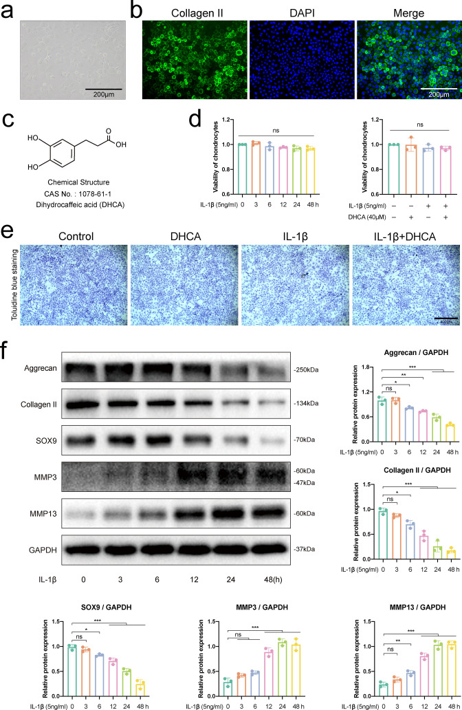
Fig. 1.
Identification of chondrocytes, detection of cell viability, and selection of proper time of interleukin-1β (IL-1β) stimulation on chondrocytes. a) Morphology of primary mouse chondrocytes under phase contrast microscope. b) Immunofluorescence analysis showed that the cytoplasm of primary mouse chondrocytes was rich in collagen II, which was stained with fluorescein isothiocyanate (FITC). c) Chemical structure of dihydrocaffeic acid (DHCA). d) Cell counting kit assesses the viability of chondrocytes treated with 5 ng/ml IL-1β for different time durations (0, 3, 6, 12, 24, and 48 hours) or with 40 μM DHCA for 24 hours. e) Toluidine blue staining of the chondrocytes treated with 40 μM DHCA or/and 5 ng/ml IL-1β for 24 hours. f) Western blot bands and quantitative analysis of mediators about anabolism (aggrecan, collagen II, and SOX9) and catabolism of chondrocytes (matrix metalloproteinase 3 (MMP3) and MMP13) in chondrocytes stimulated with 5 ng/ml IL-1β at different timepoints (0, 3, 6, 12, 24, and 48 hours). Data were presented as means and standard deviations (n = 3). GAPDH, glyceraldehyde 3-phosphate dehydrogenase; ns, no significance; *p < 0.05; **p < 0.01; ***p < 0.001.

