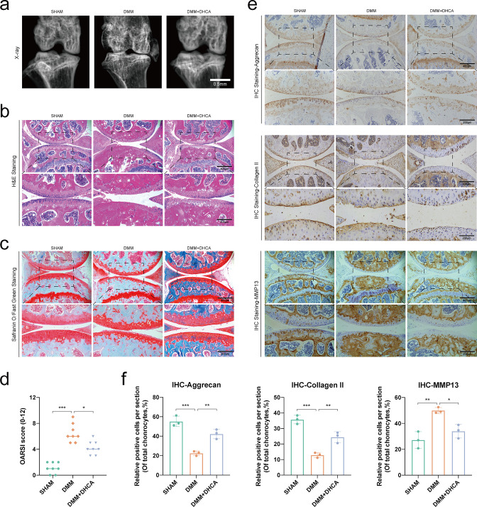
Fig. 8.
Dihydrocaffeic acid (DHCA) attenuated mouse knee cartilage degradation in vivo. a) Radiograph, b) haematoxylin and eosin (H&E) staining, and c) Safranin O/fast green staining of the knee articular cartilage among the SHAM, DMM, and DMM + DHCA group (magnification: ×100 for 400 μm, ×200 for 200 μm). d) Osteoarthritis Research Society International (OARSI) scoring analysis demonstrated the degree of cartilage degradation (n = 8). e) Immunohistochemical (IHC) staining and f) quantitative analysis of positive chondrocytes about anabolism (aggrecan and collagen II) and catabolism (MMP13) (n = 3). Data were presented as means and standard deviations. *p < 0.05; **p < 0.01; ***p < 0.001.

