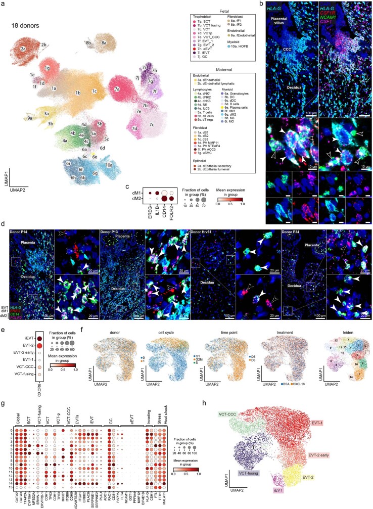
Extended Data Fig. 11. Predicted interactions between trophoblast and maternal immune cells.
a: UMAP (uniform manifold approximation and projection) scatterplot of single-cell RNA sequencing (scRNA-seq) and single-nuclei RNA sequencing (snRNA-seq) data of the 18 donors described in Extended Data Fig. 1c of the maternal-fetal interface (n = 325,665 cells and nuclei) coloured by cell state. Integration was performed with scVI. b: (Left) High-resolution imaging of a section of the placenta-decidua interface stained by smFISH for HLA-G, highlighting EVTs invading the decidua from the CCC. (Right) multiplexed co-staining with NCAM1 (dNK marker), CSF1 and cognate receptor CSF1R; dashed squares indicate areas shown magnified to right. (Bottom) solid and outlined arrows indicate neighbouring CSF1R-expressing EVTs and CSF1-expressing dNK cells, respectively. Representative image of samples from three donors. c: Dot plot showing normalised, log-transformed and variance-scaled gene expression of macrophage markers (X-axis) in data from (a) (Y-axis). d: High-resolution imaging of the placenta-decidua interface stained by multiplexed smFISH for HLA-G (EVTs), EREG (dM1), and CD14 and FOLR2 (dM2) for n = 4 donors (donor ID is specified in each panel). e: Dot plot showing normalised, log-transformed and variance-scaled expression of CXCR6 (X-axis) on the EVT subsets present in TSC (n = 2). f: UMAP scatterplots of scRNA-seq of TSC (CXCL16 and BSA conditions) coloured by donor, cell cycle phase, time point, treatment and unbiased clustering using leiden (n = 2). g: Dot plot showing normalised, log-transformed and variance-scaled expression of marker genes of the main trophoblast subsets (X-axis) in cell clusters defined in (f) (Y-axis) from the integrated manifold of CXCL16 and BSA conditions in trophoblast stem cell (TSC) scRNA-seq (n = 2). h: UMAP scatterplot of scRNA-seq of TSC coloured by cell state (n = 2). Villous cytotrophoblast (VCT), cytotrophoblast cell column (CCC), proliferative (p), extravillous trophoblast (EVT), interstitial EVTs (iEVTs), giant cells (GC), endovascular EVT (eEVT), dendritic cells (DC), lymphatic (l), maternal (m), fetal (f), Hofbauer cells (HOFB), innate lymphocytes (ILC), macrophages (M), monocytes (MO), natural killer (NK), perivascular (PV), decidual (d), epithelial (epi), stromal (S), fibroblasts (F), uterine smooth muscle cells (uSMC), bovine serum albumin (BSA).

