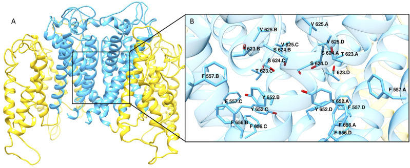
FIGURE 1.
(A) Cryo-EM structure of hERG channel in complex with astemizole (PDB ID 7CN1). The α-helixes S1-S4 constituting the voltage sensor are shown as yellow ribbons, while cyan ribbons represent the core region of the channel. (B) Close view of the main ligand binding site for hERG blockers. The residues that are reported to be mainly involved in the interactions with hERG binders are displayed as cyan sticks.

