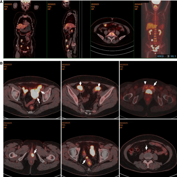
Figure 1.
Target lesions of the patient with uterine IMT by PET/CT scans. (A, B) PET/CT scans indicated multiple metastases with increased 18F-fluoro-2-deoxyglucose (18F-FDG) uptake in the vaginal stump, left-side vulva, abdominal pelvic cavity, mesentery, and iliac paravascular and retroperitoneal lymph nodes in the abdomen and pelvic cavity. IMT, inflammatory myofibroblastic tumor.

