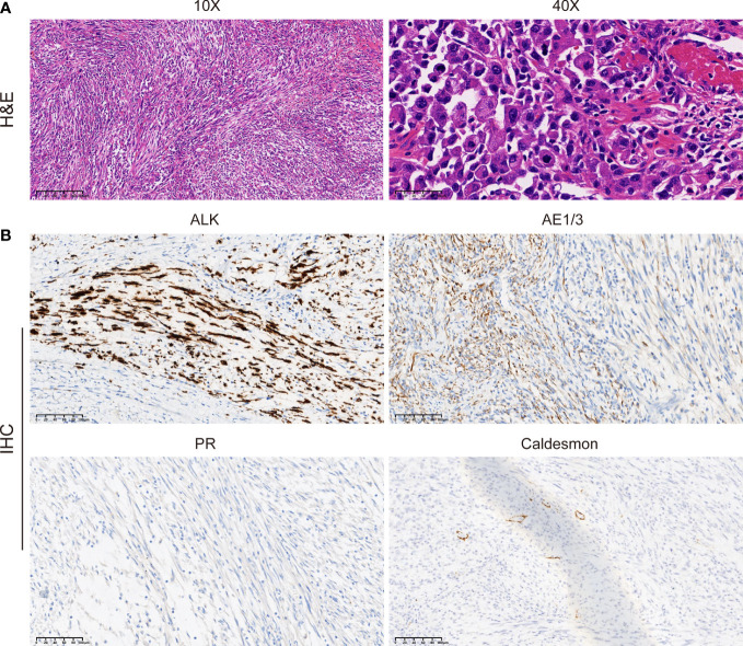
Figure 2.
Morphologic features and IHC staining of the uterine IMT samples. Representative pictures of H&E staining (A) and IHC analysis (B) of ALK, AE1/3, PR, and caldesmon expression levels in uterine IMT. Scale bars are illustrated in the pictures. IHC, immunohistochemistry; IMT, inflammatory myofibroblastic tumor; H&E, hematoxylin, and eosin.

