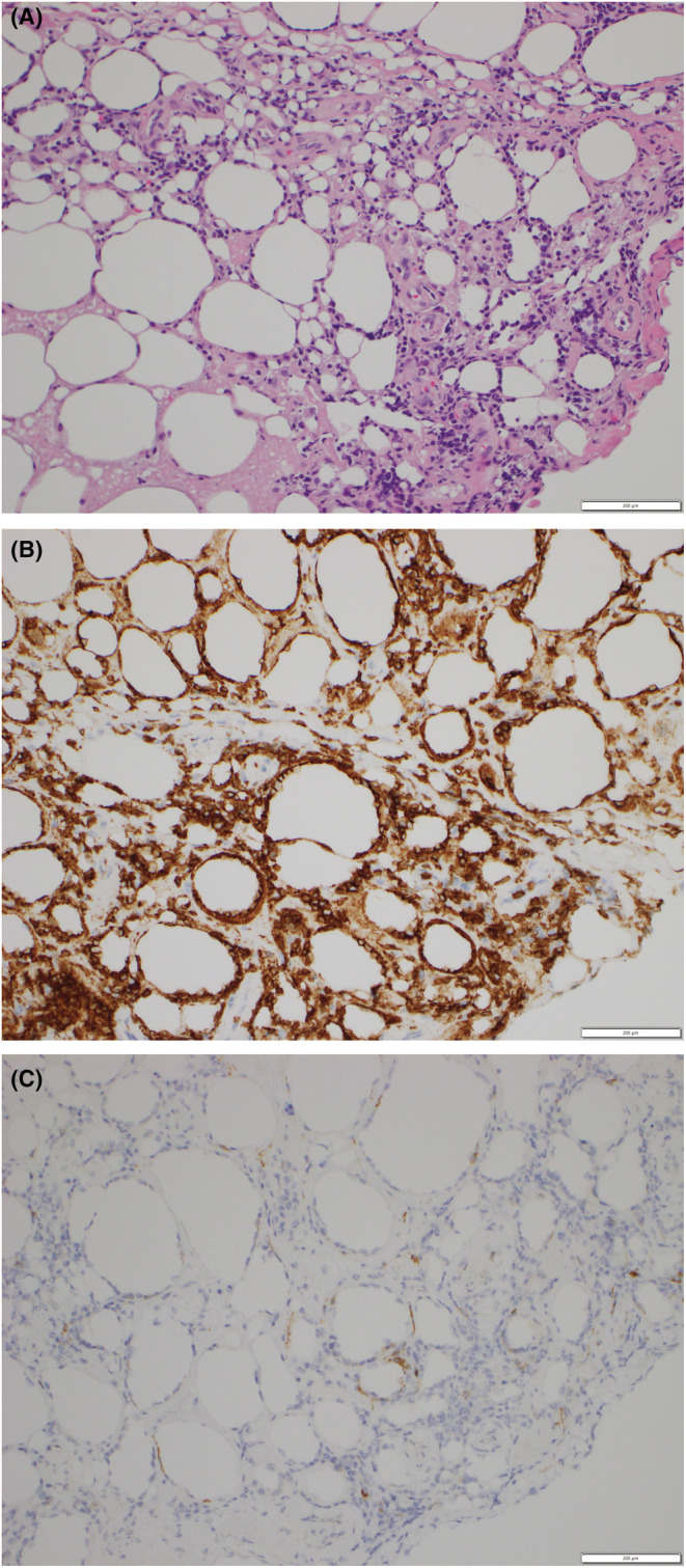
FIGURE 2.

Atypical lymphocytes and associated histiocytes that are rimming the adipocytes, in a lace‐like manner resembling panniculitis. The neoplastic infiltrate was composed of pleomorphic T cells with irregular and hyperchromatic nuclei. Hematoxylin–Eosin stain 400× (A). Immunohistology staining showed positivity for CD8 (B) and CD56 (C).
