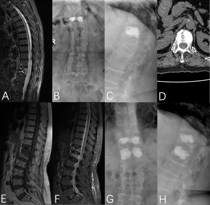
Figure 6.
A 76-year-old female was admitted to the hospital with no obvious cause of low back pain for 1 week, was diagnosed with OVCF (T12), and underwent PVP under local anesthesia on the third day after admission. (A) Preoperative fat-suppressed image showed T12 vertebral fracture, (B, C) T12 vertebral bone cement filling could be seen in the anterior and lateral X-rays after the operation, (D) Postoperative CT showed that the bone cement was unevenly distributed on the bilateral sides of the vertebrae, (E, F) MRI showed L1 vertebral fracture 5 months after the operation, (G, H) T12 and L1 vertebral bodies were filled with bone cement in the anterior and lateral X-rays after the operation.

