Fig. 3.
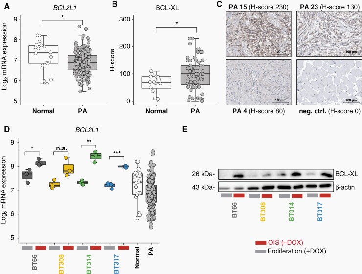
BCL-XL expression in PA. (OIS (−DOX), oncogene-induced senescence, 5 days doxycycline withdrawal; proliferation (+DOX): +1 µg/ml doxycycline). (A) BCL2L1 mRNA expression in primary PA compared to normal cerebellum. Expression data: ps_mkheidel_mkdkfz209_u133p2. Unpaired t-test: *P < .05. (B) H-scores of BCL-XL protein staining intensity in 75 primary PA samples compared to 16 inconspicuous CNS tissues adjacent to low-grade gliomas. Unpaired t-test: *P < .05. (C) Exemplary microscopic images of BCL-XL immunohistochemistry in PA showing strong (PA 15), medium (PA 23), and weak (PA 4) staining. neg. ctrl.: negative control, muscle tissue. Scale bars indicate a distance of 100 µm. (D) BCL2L1 mRNA expression in four PA cell lines in proliferation vs. OIS compared to normal cerebellum (n = 18) and primary PA (n = 191) (ps_mkheidel_mkdkfz209_u133p2). Unpaired t-test: *P < .05, **P < .01, ***P < .001. (E) Western blot of BCL-XL protein in proliferation vs. OIS mode.

