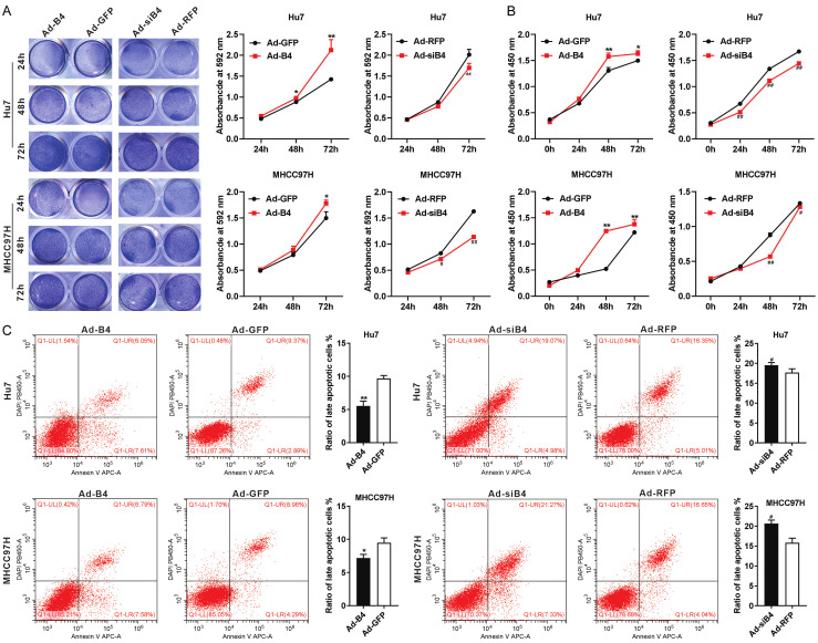
Figure 2.
BMP4 promotes proliferation and inhibits apoptosis of HCC cells under hypoxia and hypoglycemia. A. Hu7 and MHCC97H cells were infected with Ad-B4, Ad-GFP, Ad-siB4 and Ad-RFP respectively, and cultured with low glucose (LG) DMED + 100 μM CoCl2, crystal violet cell viability assay and quantitative analysis of crystal violet staining were carried out at 24 h, 48 h and 72 h. “**” P < 0.01, “*” P < 0.05, Ad-B4 group vs. Ad-GFP group, “##” P < 0.01, “#” P < 0.05, Ad-siB4 group vs. Ad-RFP group. B. Hu7 and MHCC97H cells were infected with Ad-B4, Ad-GFP, Ad-siB4 and Ad-RFP respectively, and cultured with low glucose (LG) DMED + 100 μM CoCl2, WST-1 assay was done to at 0 h, 24 h, 48 h, and 72 h. “**” P < 0.01, “*” P < 0.05, Ad-B4 group vs. Ad-GFP group, “##” P < 0.01, “#” P < 0.05, Ad-siB4 group vs. Ad-RFP group. C. Hu7 and MHCC97H cells were infected with Ad-B4, Ad-GFP, Ad-siB4 and Ad-RFP respectively, and cultured with low glucose (LG) DMED + 100 μM CoCl2, flow cytometry analysis was conducted, and the ratio of late apoptotic cells (%) was calculated at 48 h. “**” P < 0.01, “*” P < 0.05, Ad-B4 group vs. Ad-GFP group, “#” P < 0.05, Ad-siB4 group vs. Ad-RFP group.

