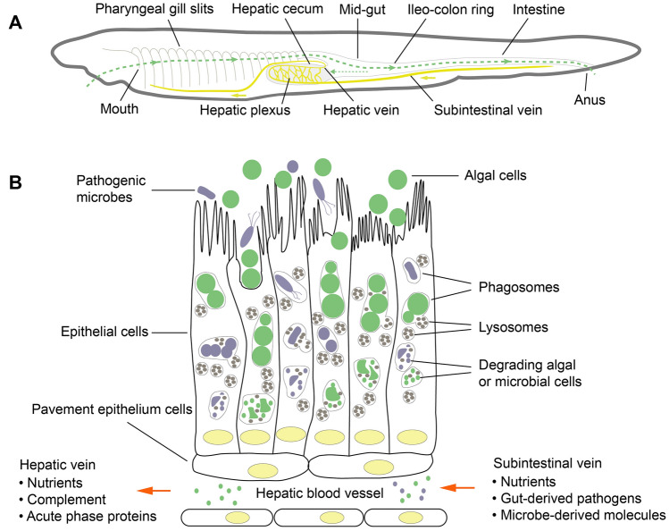
Fig. 1.
Anatomical organization of the hepatic cecum as an immune organ of amphioxus. a The digestive system, including the pharyngeal gill slits, hepatic cecum and intestine, is regarded as the major line of defense against microbial infection. When food particles enter the pharynx, they are trapped by mucus and formed into a mucus cord, which is then transported posteriorly to the mid-gut. In the mid-gut, food particles within the mucus cord are mixed with digestive enzymes, and some small particles containing algal and microbial cells (around 2 μm in diameter or less) pass into the lumen of hepatic cecum, a structure arising from the mid-gut and expands anteriorly along the right side of the pharynx. b These algal and microbial cells are phagocytized by the epithelial cells of hepatic cecum and degraded by lysosomes. Then, the degrading products enter the hepatic blood vessel plexus, in which the blood is mainly delivered from intestine via the subintestinal vein which is ultimately joined to the hepatic vein. Gathering intestine-derived blood components and hepatic epithelial cells derived products, the blood in the hepatic plexus is rich in nutrients, pathogen-derived molecules, and even surviving pathogenic microbes. The cells of the hepatic cecum are responsible for the production of complement proteins and acute phase proteins

