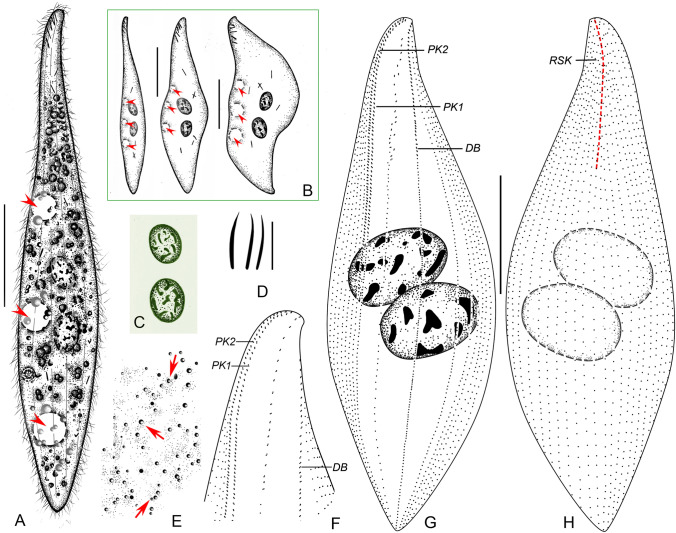
Fig. 6.
Amphileptus orientalis sp. nov. from life (A–E) and after protargol impregnation (F–H). A Left view of a representative individual, arrowheads point to the three ventral contractile vacuoles. B Shape variants, arrowheads denote the three contractile vacuoles. C Nuclear apparatus. D Oral extrusomes. E Cortical granules (arrows) of the left side. F Detail of the anterior region of the left side, showing the oral and somatic ciliary pattern. G Ciliary pattern of the left side of the holotype specimen. H Ciliary pattern of the right side of the holotype specimen, red dashed line denotes the anterior suture. DB dorsal brush, PK1 perioral kinety 1, PK2 perioral kinety 2, RSK right somatic kineties. Scale bars = 100 μm

