Fig. 7.
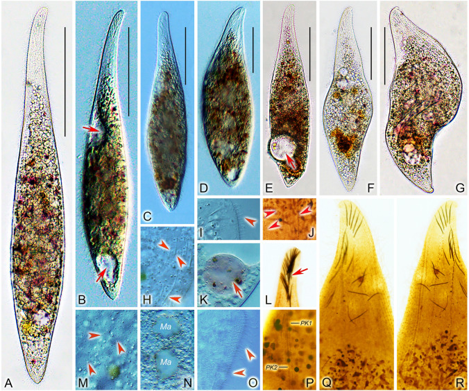
Amphileptus orientalis sp. nov. from life (A–G, H, I, K, M–O) and after protargol impregnation (J, L, P–R). A–F Left side views of extended or only slightly contracted individuals, arrows mark the ventral contractile vacuoles. G Left side view of a contracted individual. H Cytoplasmic extrusomes (arrowheads). I Lateral view, showing the dorsal brush bristles (arrowhead). J Detail showing the very loosely arranged left somatic kineties (arrowheads). K Food vacuole (arrow). L Extrusomes attached to the oral slit (arrow). M Details of cell surface, showing cortical granules (arrowheads) of the left side. N Showing the two macronuclear nodules. O Somatic cilia (arrowheads) of lateral rightmost side kineties. P Detail of the oral apparatus, showing two perioal kineties, one right and one left of the oral slit. Q, R Details of the anterior region of the right (Q) and the left (R) side of the holotype specimen, showing the ciliary and extrusome patterns. Ma macronuclear nodules. PK1 perioral kinety 1, PK2 perioral kinety 2. Scale bars = 100 μm

