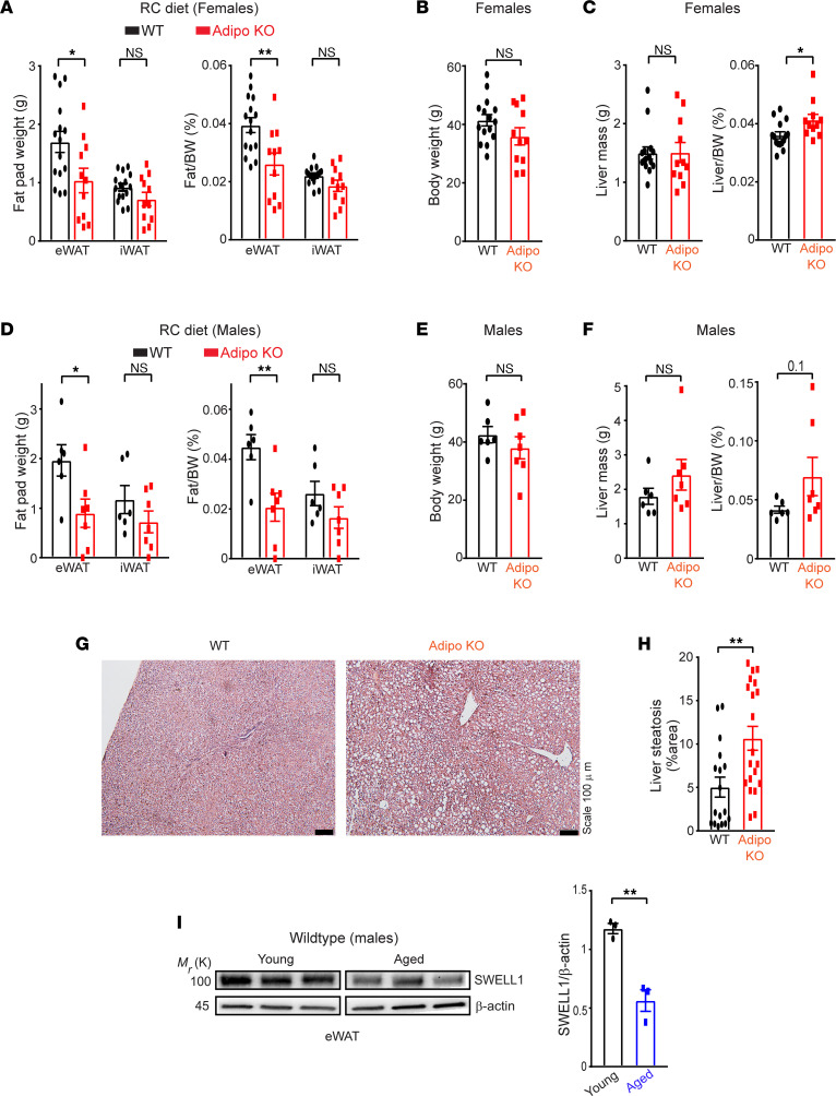
Figure 5. Adipose SWELL1 expression protects against age-related NAFLD and declines with aging.
(A) Total mass of epididymal (eWAT) and inguinal (iWAT) fat pads and their corresponding ratio of fat pad over body weight, (B) total body weight, (C) total liver mass, and ratio of liver mass over body weight dissected from WT (n = 15) and Adipo-KO (n = 11) female mice ~12 months old on RC diet. (D) Total mass of epididymal and inguinal fat pads and their corresponding ratio of fat pad over body weight, (E) total body weight, (F) total liver mass, and ratio of liver mass over body weight dissected from male WT (n = 6) and Adipo-KO (n = 7) male mice ~18–21 months old on RC diet. (G) Representative images of H&E-stained liver sections of WT and Adipo-KO mice from B (Scale bar: 100 μm). (H) Liver steatosis (%area) estimated from H&E-stained liver sections in G of WT (n = 2, 17 ROIs) and Adipo-KO (n = 2, 20 ROIs) mice using ImageJ software. (I) Representative image of Western blot comparing SWELL1 protein expression in epididymal adipose tissue isolated from young (2–3 months old, males) and aged (18–21 months old, males) WT mice fed with RC diet (left) and its corresponding densitometric ratio (right). Data are represented as mean ± SEM. Two-tailed unpaired t test was used in A–F, H, and I where *, P < 0.05, and **, P < 0.01.

