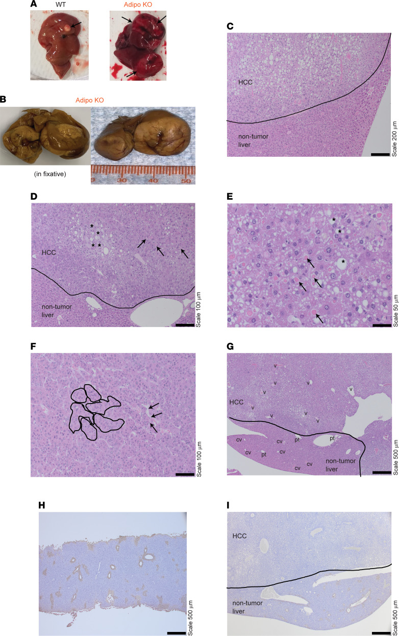
Figure 6. Male adipose SWELL1-KO mice develop HCC with aging.
(A) Representative images of livers isolated from male WT and Adipo-KO mice with tumors indicated by black arrows. (B) Formalin-fixed Adipo-KO liver with massive hepatic tumors. (C and D) H&E-stained liver sections of Adipo-KO mice with indicated nontumor and tumor-containing regions. * indicates enlarged and pleomorphic cells, and black arrows indicate eosinophilic hyaline globules in D. (E–G) Liver sections of Adipo-KO mouse with HCC morphological features. * indicates large- and intermediate-sized fat droplets, and black arrows indicate eosinophilic hyaline droplets within the HCC cells in E. Nested islands of HCC are outlined in solid black lines, and the black arrows indicate the endothelial wrapping around these nests with flattened endothelial cells in F. Abnormal veins in HCC regions are indicated with “v,” and normal portal tracts and central veins are indicated as “pt” and “cv” in the nontumorous region in G. (H and I) Representative images of glutamine synthetase staining from a normal WT (H) and Adipo-KO (I) liver with HCC. Each is a 10× eyepiece with a 4×, 10×, 20×, 40× objective; 40× magnification for images in G–I; 100× magnification for image in C; 200× magnification for images in D and F; and 400× magnification for image in E.

